.jpg)
.jpg)
-cr-390x330.jpg)
-cr-390x330.jpg)
 Fast delivery
 Training and trial
 Flexible payment terms
 Warranty
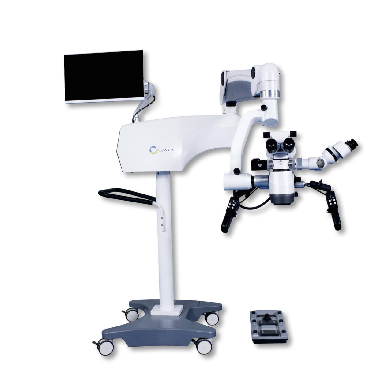
ASOM-5 surgical microscope is a high-precision instrument designed for use in various fields of surgery. Due to its characteristics, it is suitable for microsurgical manipulations, such as suturing blood vessels and nerve trunks, which makes it indispensable in neurosurgery, ENT surgery, spine surgery, orthopedics and other fields (except ophthalmology).
One of the major advantages of ASOM-5 is the ability to use ICG (indocyanine green) fluorescence to visualize the microcirculation and lymph drainage from tumors to signaling nodes. This significantly increases the accuracy of surgical interventions, reduces the risk of damage to healthy tissues and improves the overall results of operations.
The built-in microscope control unit is equipped with a 4K display and contains controls, USB ports and illumination lamps. The microscope rotates in all directions, providing maximum flexibility in positioning. The microscope allows magnification of 10× at a distance of 600 mm, 15× at 400 mm and 24× at 250 mm, which allows surgeons to work with high precision at various stages of operations.
The lighting system includes two xenon lamps with a power of 300 W each, with a brightness of 100,000 lux. If one lamp fails, a backup one is automatically activated within 10 seconds, which ensures continuity of lighting. A third spare bulb is also included.
The configuration of the microscope includes two binoculars (the main one and an assistant one according to the "face-to-face" scheme), each of which has an interpupillary distance adjustment (from 55 to 75 mm) and a diopter correction (±6 diopters). The binoculars can rotate 200 degrees with fixation, and also have rubber pads for comfortable contact with the surgeon's face.
The optics of the microscope include aspheric achromatic lenses that improve image quality due to better light collection.
You can use a 75% solution of ethyl alcohol to disinfect the external surfaces of the microscope, and 100% isopropyl alcohol to clean the optics. It is possible to use a transparent protective case that provides access to control elements.
Additional options include a third 90-degree assistant binocular, a foot pedal to control microscope functions, and a motorized drive to move the field of view.
• Magnification: 1.25× - 17×
• Working distance: 215-210 mm
• Eyepiece: 12.5× / 10×
• Distance between pupils: 55mm-75mm
• Assistant tube: Rotates 360°
• Illumination field: > 690 mm
• Image sensor type: CMOS
• Scanning method: Progressive scanning
• Frame rate: 60 fps
• Horizontal resolution: >1700 lines
• Vertical resolution: >1200 lines
• Coefficient of linear approximation: R2>0.99
• Signal-to-noise ratio: > 50 dB
• Output modes: HDMI, DVI, SDI, LVDS, RJ45, USB
• Fluorescent module: 800 nm